Advanced pharmaceutical bulletin. 12(4):828-834.
doi: 10.34172/apb.2022.084
Research Article
Decreased Expression of LAMB3 Is Associated with Esophageal Cancer Stem Cell Formation
Anoosheh Ehtesham 1  , Ayyoob Khosravi 2, 3 , Marie Saghaeian Jazi 2 , Jahanbakhsh Asadi 1, 2, * , Seyyed Mehdi Jafari 1, 4, *
Author information:
1Metabolic Disorders Research Center, Golestan University of Medical Sciences, Gorgan, Iran.
2Stem Cell Research Center, Golestan University of Medical Sciences, Gorgan, Iran.
3Department of Molecular Medicine, Faculty of Advanced Medical Technologies, Golestan University of Medical Sciences, Gorgan, Iran.
4Department of Biochemistry and Biophysics, School of Medicine, Golestan University of Medical Sciences, Gorgan, Iran.
Abstract
Purpose:
Esophageal squamous cell carcinoma (ESCC) is a highly aggressive cancer. The main cause of death in ESCC is related to relapse, metastasis, and resistance to cancer therapy. Recent studies have shown that a minor subset of cancer cells, known as cancer stem cells (CSCs), are responsible for tumor formation initiation and cancer progression. Understanding the genes associated with CSCs and metastasis can help in targeted cancer therapy. The aim of this study was to assess the expression of LAMB3 and TOP2A metastasis-associated genes in CSCs and adherent cells in the xenograft mouse model.
Methods:
Esophageal CSCs were enriched by the sphere formation method. The expression level of LAMB3 and TOP2A genes were evaluated in spheres and adherent cells in vitro by qRT-PCR. A xenograft mouse model was established to investigate the tumorigenesis and metastasis potential by subcutaneous and tail vein injection of CSCs and adherent YM-1 cells. Consequently, LAMB3 and TOP2A expression at the mRNA level was assessed in tumors. Immunohistochemistry was also used to evaluate the LAMB3 expression at the protein level in tumors.
Results:
CSCs-derived tumor was developed more quickly than the adherent cells-derived tumor. LAMB3 at mRNA and protein level was significantly down-regulated in sphere-derived tumor compared with adherent cells-derived tumor (P value <0.05). TOP2A expression was almost similar in both sphere cells and adherent cells and there was no significant difference.
Conclusion:
we concluded that YM-1 spheres have CSCs characteristics in vitro with high capability of tumorigenicity in vivo. Our results were also shown that the LAMB3 expression was decreased in YM-1 spheres suggesting LAMB3 association with sphere formation.
Keywords: Esophagus cancer, Cancer stem cell, LAMB3 protein, Topoisomerase II alpha
Copyright and License Information
©2022 The Authors.
This is an Open Access article distributed under the terms of the Creative Commons Attribution (CC BY), which permits unrestricted use, distribution, and reproduction in any medium, as long as the original authors and source are cited. No permission is required from the authors or the publishers.
Introduction
Esophageal cancer (EC) is the eighth among the most common malignancies and the sixth cause of death from cancer worldwide.
1
Esophageal squamous cell carcinoma (ESCC), a major subtype of EC, is considered as highly aggressive malignancy of the digestive tract.
2
Despite recent advances in radiotherapy, chemotherapy and surgical therapeutic approaches, the 5-year survival rate of ESCC patients is still less than 15%.
3
Recently, studies have shown that a limited subset of cancer cells, known as cancer stem cells (CSCs) or tumor-initiating cells play a critical role in tumorigenicity and metastasis.
4
The CSCs, similar to normal stem cells, are able to self-renew and have differentiation potential to different cell lineages.
4
The CSCs take part important function in tumorigenesis and even low numbers of them are able to initiate primary tumor formation and metastasis progression.
5
These small subpopulation have been already identified in many solid tumors
4
including EC.
6
Considerably, Gene expression profile in CSCs is exclusive and necessary to initiate and progress tumors and metastasis.
7
Different metastasis-associated genes are playing important roles in high invasive ability of CSCs. LAMB3 encodes β3, one of the three chains of Laminin-332, which have various functions in invasion, migration and progression of metastasis in different cancers.
8
The MMP-7 can produce a cleaved fragment of 90 kD originated from LAMB3 which can induce the migration of colon carcinoma cells.
9
Moreover, during the pulmonary metastasis early arrest of tumor cells happens through the interaction of α3β1 integrin with LN-332.
10
The second gene in our study is TOP2A that encodes 170kD topoisomerase IIa (topoIIa) which is involved in DNA synthesis, gene transcription and DNA topology.
11,12
It plays a critical role in development, proliferation, and invasion in carcinomas.
13,14
Interestingly, dysregulation of TOP2A is significantly associated with tumor proliferation and progression such as, pancreatic
15
and EC.
16
Recent studies have more evaluated LAMB3 and TOP2A expression in malignant tumors rather than in CSCS. Studies have shown that LAMB3 and TOP2A were overexpressed in malignant tissues compared with normal tissues in ESCC.
17
In the current study, we aimed to investigate the expression of these two metastasis-associated genes in CSCs of ESCC in vitro and in vivo.
Materials and Methods
Cell culture
Human ESCC YM-1 cell line has already been established in our laboratory at Golestan University of Medical Sciences.
18
Cells were cultured as a monolayer in high glucose DMEM/F12 (BioIdea, CN:1027) supplemented with 10% fetal bovine serum (FBS) (BioIdea, CN:10270-106), 100 IU/mL penicillin and 100 μg/mL streptomycin (BioIdea, CN:1036) at 37°C in a humidified 5% CO2 incubator. The medium was changed every 3 days and cells were passaged to another flask every week.
Sphere-forming assay
To obtain sphere cultures, monolayer cells in DMEM/F12 medium containing FBS 5% were washed with PBS (BioIdea). The cells were dissociated using Trypsin-EDTA 0.25% (BioIdea, CN:1002). Subsequently, the dissociated cells were seeded at 20,000 cells/mL in the low attachment petri dish (Labtrone, Iran). In petri dishes, the cells were suspended in serum-free DMEM/F12 supplemented with antibiotics, 20 ng/mL of human epidermal growth factor (EGF; ROYAN, CN:1102-25), 20 ng/mL of human basic fibroblast growth factor (bFGF; ROYAN, CN:1101-25), and 2% B-27 50x supplement (GIBCO, CN:17504-044).
Sphere passaging
Spheres were passaged up to five times every 3-4 days. The process of sphere passaging takes approximately 15 to 20 days. The spheres were collected from petri dish gently, then dissociated with trypsin-EDTA. The single cells were then centrifuged and re-suspended in a new petri dish containing serum-free medium and growth factors. The number and volume of spheres were counted by Trypan blue staining under an optical microscope.
Subcutaneous xenograft mouse model
YM-1 parental and sphere-forming cells were divided into two groups. The number of 106 single cells in PBS 100 μL/Matrigel 100 μL (1:1) mixture were subcutaneously injected into 4week-old male athymic nude mice weighing 16-18 grams. The nude mice were maintained under specific pathogen-free conditions in Pasteur Institute, Amol, Iran. When the tumor size achieved to minimum 1 cm in diameter, mice were sacrificed. The primary tumors were harvested and minced into small pieces. Part of the tissue was used for the molecular experiment and others were fixed in 10% formalin then embedded in paraffin for histological and immunohistological staining. To ensure the tumor pathology, the H&E staining was performed in the 5 Azar hospital laboratory, Gorgan, Iran.
Experimental metastatic xenograft mouse model
To carry out the experimental metastatic mouse model, cells were injected into the nude mice via the lateral tail vein and intraperitoneal. Sphere cells in PBS (106 cells per mouse) and adherent cells in PBS (106, 3×10⁶ and 5×10⁶ cells per mouse) were used in separate groups. Moreover, for more assurance, intraperitoneal injection also was checked for metastasis experiments (106 cells per mouse). Animals were euthanized when the moribund signs were observed in them or after 3 months of injection if they survived. Lungs, liver, femur, and bone were isolated and fixed in 10% formalin for hematoxylin and eosin (H&E) staining. The stained sections were observed under the microscope.
RNA extraction, reverse transcription and quantitative real-time PCR ( qRT -PCR)
Total RNA was isolated from cells and tumors using TRIZOL (Invitrogen CN:15596-026). The first strand cDNA was synthesized using a reverse transcriptase kit (Yekta tajhiz) following the manufacturer’s protocol. Then qPCR was performed using SYBR Green PCR Master Mix kit (Applied Biosystems, CN:4367659) by ABI 7300 Real time PCR instrument. The GAPDH gene was used as housekeeping control to normalize gene expression level. GAPDH, LAMB3 and TOP2A were amplified using the following thermal condition: 95°C for 5 minutes, 38 cycles of 95°C for 10 seconds, 60°C for 30 seconds and 72°C for 40 seconds following with melt curve analysis and thermal condition for SOX2 and NANOG was 95°C for 2 minutes, 38 cycles of 95°C for 15 seconds, 62°C for 1 minutes and 72°C for 1.30 minutes and final extension 72°C for 10 minutes. Expression of genes was measured using 2−ΔCt. Experiments were performed in triplicate.
The sequence of used specific primers were as following: LAMB3 forward: 5’-GTA TGG CGA GTG GCA GAT GA-3’ and reverse: 5’-GCA GAG AGA CAG GGT TCA CA-3’, TOP2A forward: 5’-GCA TTC CTA CAT CCA AGG GTG G-3’ and reverse: 5’-TGT CTG AGA GTC AAA GGT TGG GTT-3’, GAPDH forward: 5’-AAG CTC ATT TCC TGG TAT GAC AAC-3’and reverse: 5’-CTC TCT TCC TCT TGT GCT CTT G -3’, SOX2 forward: TACAGCATGTCCTACTCGCAG and reverse: GAGGAAGAGGTAACCACAGGG and NANOG forward: ATTCAGGACAGCCCTGATTCTTC and reverse: TTTTTGCGACACTTCTCTGC
Immunohistochemistry (IHC)
To assess the LAMB3 protein expression at tissue sections, the fixed xenograft tumor sections from adherent and sphere cells were stained using the anti-LABM3 antibody. Formalin-fixed and paraffin-embedded tumor sections with 5 µm thickness were used for staining. The sections were deparaffinized with xylene and then were rehydrated with ethanol. Antigen retrieval was used to break the possible methylene bridges. The sections were washed with buffer and then were incubated with 3% H2O2 and 1% bovine serum albumin (BSA) respectively. Slides were then placed in a humid chamber. Slides were stained with the primary mouse monoclonal antibody against LAMB3 (Santa Cruz, sc-133178) diluted 1:50 for overnight. Subsequently, slides were washed with buffer and incubated with a mouse IgGk protein conjugated to horseradish peroxidase (m- IgGk-HRP, sc-516102). The stained xenograft tumors tissue sections were then incubated with DAB solution. Finally, the slides were counterstained by hematoxylin and were studied using light microscopy.
Statistical analysis
Two-tail Student’s t test and ANOVA were used to analyze differences between groups. In the case of non-normal distribution, the non-parametric test was used. All statistical analysis was performed by SPSS 16.0 software. P-values of less than 0.05 were recognized as statistically significant. The graphs were drawn by GraphPad Prism v 5.04.
Results and Discussion
The enriched YM-1 spheres show CSCs characteristics
CSCs are tumor initiating-cells that are able to generate heterogeneous cell population within a tumor.
19
In a cancer cell line, CSCs can be identified with different features including sphere formation assay, over-expression of stemness genes, increased drug resistance and high in vivo tumorigenesis potential.
20,21
In sphere forming-assay, CSCs subpopulation can be enriched and have self-renewal and independent growth in vitro.
21,22
In this study, to evaluate the presence of CSCs, we isolated CSCs from YM-1 EC cells with sphere formation assay. YM-1 adherent cells were cultured in serum-free, low attachment three-dimensional (3D) culture medium. Subsequently only a limited number of cells were able to grow and self-renew (Figure 1A). In this condition, single cells formed sphere clusters as shown in (Figure 1B and 1C).
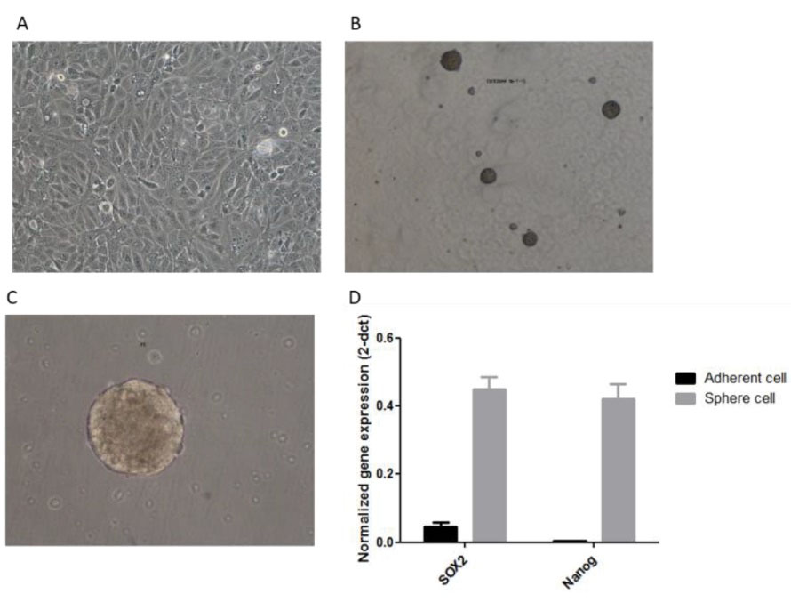
Figure 1.
YM-1 cells in special conditions represent features of CSCs.(A) adherent cells in DMEM/F12 medium supplemented with 10% FBS. (B and C) Sphere clusters in specific 3D culture (×4 and ×40). (D) mRNA level of SOX2 and NANOG are up-regulated in spheres vs adherent cells.Bars represent the normalized gene expression (2-dct) with standard error bars
.
YM-1 cells in special conditions represent features of CSCs.(A) adherent cells in DMEM/F12 medium supplemented with 10% FBS. (B and C) Sphere clusters in specific 3D culture (×4 and ×40). (D) mRNA level of SOX2 and NANOG are up-regulated in spheres vs adherent cells.Bars represent the normalized gene expression (2-dct) with standard error bars
The spheres were checked for stemness gene expression and as shown in (Figure 1D), significant overexpression of the SOX2 and Nanog was observed in YM-1 sphere cells. The sphere formation capability of the YM-1 cells was shown previously in different projects in our lab but checked again in the current study.
Sphere-forming cells exhibit higher tumorigenicity compared with parental cells in vivo
For more confirmation that the YM-1 spheres have CSCs features, we aimed to develop xenograft mouse model with subcutaneous injection. The results indicated 106 enriched YM-1 spheres formed a tumor measuring 1 cm in diameter after one month while 106 adherent cells formed a tumor in this size after two months (Figure 2A). The histology of both tumors was confirmed as ESCC tumor by (H&E) staining (Figure 2B). At the end, our findings confirmed that the spheres subpopulation of the YM-1 EC cells harbors most of the characteristics of CSCs.
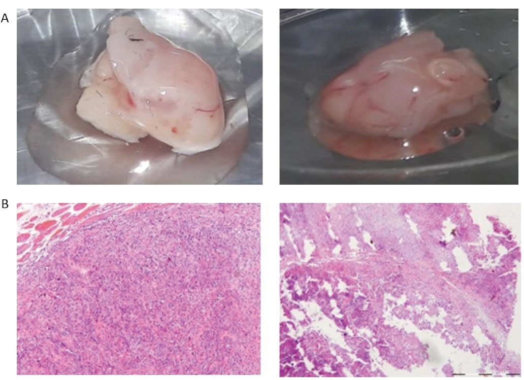
Figure 2.
The high tumorgenicity ability of spheres. (A) The spheres-derived tumor after only a month reached the same tumor size (1 cm in diameter) of adherent cells-derived tumor following 2 months. (B) The H&E staining of xenograft tumors confirmed the neoplastic histology of the tumors (×4)
.
The high tumorgenicity ability of spheres. (A) The spheres-derived tumor after only a month reached the same tumor size (1 cm in diameter) of adherent cells-derived tumor following 2 months. (B) The H&E staining of xenograft tumors confirmed the neoplastic histology of the tumors (×4)
YM-1 cell line metastatic potential
To assess the metastatic potential of ESCC YM-1cell line, we developed a xenograft mouse model by experimental metastatic assay in which cells were injected directly into the systemic circulation.
23
Mice were injected with YM-1 sphere or adherent cells via a tail vein and intraperitoneal injection. Then they were sacrificed following maximum 3 months follow up and all of the organs were isolated. Since no nodule was found we stained lungs, liver, femur, and bone of the mice with H&E staining to make a more accurate assessment. There was no indication of metastasis under the microscope (data not shown). Due to our results, we conclude that probably YM-1 cell line or spheres have poor ability to form secondary metastatic tumors which could be explained with the cell-specific characteristics.
LAMB3 expression assessment at mRNA level in spheres and adherent cells
Interaction of CSCs with their microenvironment can lead to cancer development and metastasis. extracellular matrix (ECM) is a major structure of CSCs niche that its protein components could be dysregulated in pathological states and cancer.
24
LAMB3 by coding β3 is involved in production of Laminin-332 as a ECM protein. Laminin-332 is made up of three subunits (α3, β3 and γ2). Studies have shown each of Laminin-332 subunits lonely, either as a dimer or as a trimer plays role in stimulating invasion, migration and homing in metastasis of colon,
9
pulmonary cancers
10
and esophagus squamous cell carcinoma.
25
However, different studies reported either up-regulation or down-regulation of Laminin-332 heterotrimer in different cancers.
26
Upregulation of LAMB3 is correlated with the depth invasion and metastasis risk of some malignant tumors such as the colorectal
27
and gastric.
8
Up to now, limited studies have examined the expression of LAMB3 and Laminin-332 in CSCs while in current study, we compared the expression of LAMB3 in YM-1spheres with CSCs features withadherent cells in vitro and in vivo by qRT-PCR method. As shown in (Figure 3B) the gene expression of LAMB3 was decreased in spheres compared with adherent cells in vitro, however, it was not significant (P value > 0.05). To investigate LAMB3 expression in vivo, we evaluated the expression at mRNA level in spheres-derived tumor and adherent cells-derived tumor in subcutaneous injection (primary tumor). Similar to in vitro results, LAMB3 expression in the spheres-derived tumor was decreased significantly (P value = 0.03) (Figure 3).
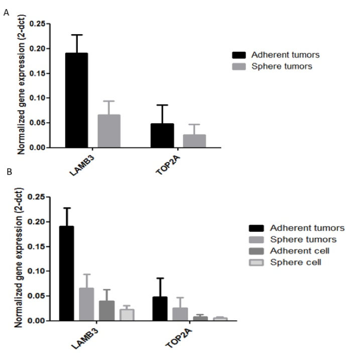
Figure 3.
Down-regulation of LAMB3 and TOP2A in spheres vs adherent cells.(A and B) qRT-PCR analysis indicated Down-regulation of LAMB3 and TOP2A in spheres compared with adherent cells in vivo and in vitro respectively
.
Down-regulation of LAMB3 and TOP2A in spheres vs adherent cells.(A and B) qRT-PCR analysis indicated Down-regulation of LAMB3 and TOP2A in spheres compared with adherent cells in vivo and in vitro respectively
Xenograft tumor derived from spheres express less LAMB3 proteins
Whereas there was a meaningful difference in LAMB3 mRNA expression between two tumors, we evaluated LAMB3 expression in protein level in two different tumors by IHC. Our observations represented that LAMB3 protein is detectable in both spheres-derived tumor and adherent cells-derived tumor. The spheres-derived tumor expresses less LAMB3 protein level in comparison to the adherent cells-derived tumor (Figure 4), which is fallowing the qRT-PCR results.
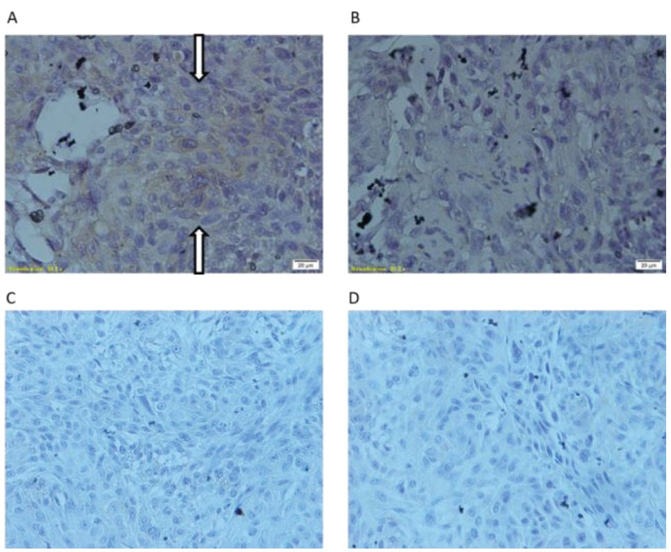
Figure 4.
Comparison of the protein expression level of LAMB3 in spheres and adherent cells in vivo.(A) Immunohistochemistry (IHC) analysis demonstrated significant down-regulation of LAMB3 in spheres-derived tumor vs adherent cells-derived tumor. (B) Up-regulation of LAMB3 expression at the protein level in adherent cells-derived tumor is showed with flash. (C and D) negative control samples
.
Comparison of the protein expression level of LAMB3 in spheres and adherent cells in vivo.(A) Immunohistochemistry (IHC) analysis demonstrated significant down-regulation of LAMB3 in spheres-derived tumor vs adherent cells-derived tumor. (B) Up-regulation of LAMB3 expression at the protein level in adherent cells-derived tumor is showed with flash. (C and D) negative control samples
Oktem et al
28
reported that LAMB3 expression is significantly upregulated in CD133+/CD44+CSCs grown as a monolayer comparing with CD133-/CD44-counterpart. Govaere et al
29
showed the role of Laminin-332 in sustaining CSCs quiescence in hepatic carcinoma. Liu et al
30
indicated that Laminin-332 promotes tumor metastasis and have a prominent role in the progression of lung cancer. In the study of Liu et al,
30
Laminin-332 was up-regulated in tumor spheres compared to adherent cells, consequently, only tumor spheres resulted in lung metastasis via tail vein injection. However, our data showed that the tumors derived from sphere cells with higher tumorigenic features express less LAMB3, both at mRNA and protein level that this significant down-regulation in expression shows relation and influence of LAMB3 insphere formation andEC progression. concordantly, Lathia et al
31
showed that LAMA2 expression is downregulated in CSC of glioblastoma and LAMA2 is identified as a malignant marker in aggressive ependymoma.
TOP2A expression assessment at mRNA level in spheres and adherent cells
Another gene studied in current study was TOP2A, the alpha subunit of the topoisomerase II which is the target of cancer therapy drugs such as anthracyclines.
32
Inhibition of TOP2A suppresses metastatic spread of tumors.
15
TOP2A expression have been rarely investigated in CSCs or tumor spheres and more commonly evaluated in cancer tumors in which TOP2A expression was correlated with advanced stages of the disease.
33
In this study, we also evaluated TOP2A expression at mRNA level in spheres and adherent cells in vitro and in subcutaneous tumors. Our findings showed non-significant downregulation of the TOP2A in spheres of both in vitro and in vivo experiments compared with adherent cells (Figure 3).However, previous studies reported TOP2A association with tumor proliferation and progression in several types of cancers such as pancreatic,
15
prostate,
33
and ESCC.
16
Terashima et al
34
indicated that TOP2A expression markedly increases the risk of hematogenous recurrence and peritoneum recurrences in gastric carcinoma. Li et al
33
illustrated that TOP2Ahigh cells related to recurrence/ metastasis but unexpectedly TOP2Aneg cells show CSCs properties in prostate cancer which is in concordant to the decreased expression of TOP2A in esophageal CSCs observed in our findings.
Conclusion
In conclusion, our results illustrated that YM-1 esophageal cancer cell spheres have CSCs characteristics in vitro with high capability of tumorigenicity in vivo. We found that the LAMB3 gene and protein expression is decreased in YM-1 spheres suggesting LAMB3 association with sphere formation and esophageal CSC development; however the underlying mechanism remains to be investigated in future experiments.
Acknowledgments
This study was supported by the Metabolic Disorders Research Centre, Golestan University of Medical Sciences (No.960413075).
Ethical Issues
In this project, nude mice were kept and treated at Pasteur Amol institute according to health instructions and it was approved in local ethic committee (No: ir.goum.rec.1395.276).
Conflict of Interest
All authors declare no conflicts of interest in this work.
References
- Ferlay J, Soerjomataram I, Dikshit R, Eser S, Mathers C, Rebelo M. Cancer incidence and mortality worldwide: sources, methods and major patterns in GLOBOCAN 2012. Int J Cancer 2015; 136(5):E359-86. doi: 10.1002/ijc.29210 [Crossref] [ Google Scholar]
- Song Y, Li L, Ou Y, Gao Z, Li E, Li X. Identification of genomic alterations in oesophageal squamous cell cancer. Nature 2014; 509(7498):91-5. doi: 10.1038/nature13176 [Crossref] [ Google Scholar]
- Lambert R. Endoscopy in screening for digestive cancer. World J Gastrointest Endosc 2012; 4(12):518-25. doi: 10.4253/wjge.v4.i12.518 [Crossref] [ Google Scholar]
-
Noll JE, Vandyke K, Zannettino AC. The role of the “cancer stem cell niche” in cancer initiation and progression. In: Wislet S, ed. Adult Stem Cell Niches. IntechOpen; 2014. p. 291-320. 10.5772/58598.
- Nimmakayala RK, Batra SK, Ponnusamy MP. Unraveling the journey of cancer stem cells from origin to metastasis. Biochim Biophys Acta Rev Cancer 2019; 1871(1):50-63. doi: 10.1016/j.bbcan.2018.10.006 [Crossref] [ Google Scholar]
- Huang D, Gao Q, Guo L, Zhang C, Jiang W, Li H. Isolation and identification of cancer stem-like cells in esophageal carcinoma cell lines. Stem Cells Dev 2009; 18(3):465-73. doi: 10.1089/scd.2008.0033 [Crossref] [ Google Scholar]
- Li F, Tiede B, Massagué J, Kang Y. Beyond tumorigenesis: cancer stem cells in metastasis. Cell Res 2007; 17(1):3-14. doi: 10.1038/sj.cr.7310118 [Crossref] [ Google Scholar]
- Ii M, Yamamoto H, Taniguchi H, Adachi Y, Nakazawa M, Ohashi H. Co-expression of laminin β3 and γ2 chains and epigenetic inactivation of laminin α3 chain in gastric cancer. Int J Oncol 2011; 39(3):593-9. doi: 10.3892/ijo.2011.1048 [Crossref] [ Google Scholar]
- Remy L, Trespeuch C, Bachy S, Scoazec JY, Rousselle P. Matrilysin 1 influences colon carcinoma cell migration by cleavage of the laminin-5 beta3 chain. Cancer Res 2006; 66(23):11228-37. doi: 10.1158/0008-5472.can-06-1187 [Crossref] [ Google Scholar]
- Wang H, Fu W, Im JH, Zhou Z, Santoro SA, Iyer V. Tumor cell alpha3beta1 integrin and vascular laminin-5 mediate pulmonary arrest and metastasis. J Cell Biol 2004; 164(6):935-41. doi: 10.1083/jcb.200309112 [Crossref] [ Google Scholar]
- Tsavaris N, Lazaris A, Kosmas C, Gouveris P, Kavantzas N, Kopterides P. Topoisomerase I and IIalpha protein expression in primary colorectal cancer and recurrences following 5-fluorouracil-based adjuvant chemotherapy. Cancer Chemother Pharmacol 2009; 64(2):391-8. doi: 10.1007/s00280-008-0886-4 [Crossref] [ Google Scholar]
- Żaczek A, Markiewicz A, Jaśkiewicz J, Pieńkowski T, Rhone P, Jassem J. Clinical evaluation of developed PCR-based method with hydrolysis probes for TOP2A copy number evaluation in breast cancer samples. Clin Biochem 2010; 43(10-11):891-8. doi: 10.1016/j.clinbiochem.2010.04.060 [Crossref] [ Google Scholar]
- Di Leo A, Desmedt C, Bartlett JM, Piette F, Ejlertsen B, Pritchard KI. HER2 and TOP2A as predictive markers for anthracycline-containing chemotherapy regimens as adjuvant treatment of breast cancer: a meta-analysis of individual patient data. Lancet Oncol 2011; 12(12):1134-42. doi: 10.1016/s1470-2045(11)70231-5 [Crossref] [ Google Scholar]
- Zhang R, Xu J, Zhao J, Bai JH. Proliferation and invasion of colon cancer cells are suppressed by knockdown of TOP2A. J Cell Biochem 2018; 119(9):7256-63. doi: 10.1002/jcb.26916 [Crossref] [ Google Scholar]
- Pei YF, Yin XM, Liu XQ. TOP2A induces malignant character of pancreatic cancer through activating β-catenin signaling pathway. Biochim Biophys Acta Mol Basis Dis 2018; 1864(1):197-207. doi: 10.1016/j.bbadis.2017.10.019 [Crossref] [ Google Scholar]
- Yu Y, Ding S, Liang Y, Zheng Y, Li W, Yang L. Expression of ERCC1, TYMS, TUBB3, RRM1 and TOP2A in patients with esophageal squamous cell carcinoma: a hierarchical clustering analysis. Exp Ther Med 2014; 7(6):1578-82. doi: 10.3892/etm.2014.1659 [Crossref] [ Google Scholar]
- Su H, Hu N, Yang HH, Wang C, Takikita M, Wang QH. Global gene expression profiling and validation in esophageal squamous cell carcinoma and its association with clinical phenotypes. Clin Cancer Res 2011; 17(9):2955-66. doi: 10.1158/1078-0432.ccr-10-2724 [Crossref] [ Google Scholar]
- Ayyoob K, Masoud K, Vahideh K, Jahanbakhsh A. Authentication of newly established human esophageal squamous cell carcinoma cell line (YM-1) using short tandem repeat (STR) profiling method. Tumour Biol 2016; 37(3):3197-204. doi: 10.1007/s13277-015-4133-4 [Crossref] [ Google Scholar]
- Baccelli I, Trumpp A. The evolving concept of cancer and metastasis stem cells. J Cell Biol 2012; 198(3):281-93. doi: 10.1083/jcb.201202014 [Crossref] [ Google Scholar]
- Schatton T, Frank NY, Frank MH. Identification and targeting of cancer stem cells. Bioessays 2009; 31(10):1038-49. doi: 10.1002/bies.200900058 [Crossref] [ Google Scholar]
- Pastrana E, Silva-Vargas V, Doetsch F. Eyes wide open: a critical review of sphere-formation as an assay for stem cells. Cell Stem Cell 2011; 8(5):486-98. doi: 10.1016/j.stem.2011.04.007 [Crossref] [ Google Scholar]
- Cao L, Zhou Y, Zhai B, Liao J, Xu W, Zhang R. Sphere-forming cell subpopulations with cancer stem cell properties in human hepatoma cell lines. BMC Gastroenterol 2011; 11:71. doi: 10.1186/1471-230x-11-71 [Crossref] [ Google Scholar]
- Khanna C, Hunter K. Modeling metastasis in vivo. Carcinogenesis 2005; 26(3):513-23. doi: 10.1093/carcin/bgh261 [Crossref] [ Google Scholar]
- Nallanthighal S, Heiserman JP, Cheon DJ. The role of the extracellular matrix in cancer stemness. Front Cell Dev Biol 2019; 7:86. doi: 10.3389/fcell.2019.00086 [Crossref] [ Google Scholar]
- Kita Y, Mimori K, Tanaka F, Matsumoto T, Haraguchi N, Ishikawa K. Clinical significance of LAMB3 and COL7A1 mRNA in esophageal squamous cell carcinoma. Eur J Surg Oncol 2009; 35(1):52-8. doi: 10.1016/j.ejso.2008.01.025 [Crossref] [ Google Scholar]
- Tsuruta D, Kobayashi H, Imanishi H, Sugawara K, Ishii M, Jones JC. Laminin-332-integrin interaction: a target for cancer therapy?. Curr Med Chem 2008; 15(20):1968-75. doi: 10.2174/092986708785132834 [Crossref] [ Google Scholar]
- Volpi A, D’Elia G, Pannarale OC, Di Gennaro F, Guida P, Martinelli E. [Overexpression of laminine-5 (LN-5) in peritoneal lavage of colorectal cancer patients preliminary results]. G Chir 2011; 32(1-2):59-63. [ Google Scholar]
- Oktem G, Sercan O, Guven U, Uslu R, Uysal A, Goksel G. Cancer stem cell differentiation: TGFβ1 and versican may trigger molecules for the organization of tumor spheroids. Oncol Rep 2014; 32(2):641-9. doi: 10.3892/or.2014.3252 [Crossref] [ Google Scholar]
- Govaere O, Wouters J, Petz M, Vandewynckel YP, Van den Eynde K, Van den Broeck A. Laminin-332 sustains chemoresistance and quiescence as part of the human hepatic cancer stem cell niche. J Hepatol 2016; 64(3):609-17. doi: 10.1016/j.jhep.2015.11.011 [Crossref] [ Google Scholar]
- Liu CC, Lin JH, Hsu TW, Hsu JW, Chang JW, Su K. Collagen XVII/laminin-5 activates epithelial-to-mesenchymal transition and is associated with poor prognosis in lung cancer. Oncotarget 2018; 9(2):1656-72. doi: 10.18632/oncotarget.11208 [Crossref] [ Google Scholar]
- Lathia JD, Li M, Hall PE, Gallagher J, Hale JS, Wu Q. Laminin alpha 2 enables glioblastoma stem cell growth. Ann Neurol 2012; 72(5):766-78. doi: 10.1002/ana.23674 [Crossref] [ Google Scholar]
- Panvichian R, Tantiwetrueangdet A, Angkathunyakul N, Leelaudomlipi S. TOP2A amplification and overexpression in hepatocellular carcinoma tissues. Biomed Res Int 2015; 2015:381602. doi: 10.1155/2015/381602 [Crossref] [ Google Scholar]
- Li X, Liu Y, Chen W, Fang Y, Xu H, Zhu HH. TOP2Ahigh is the phenotype of recurrence and metastasis whereas TOP2Aneg cells represent cancer stem cells in prostate cancer. Oncotarget 2014; 5(19):9498-513. doi: 10.18632/oncotarget.2411 [Crossref] [ Google Scholar]
- Terashima M, Ichikawa W, Ochiai A, Kitada K, Kurahashi I, Sakuramoto S. TOP2A, GGH, and PECAM1 are associated with hematogenous, lymph node, and peritoneal recurrence in stage II/III gastric cancer patients enrolled in the ACTS-GC study. Oncotarget 2017; 8(34):57574-82. doi: 10.18632/oncotarget.15895 [Crossref] [ Google Scholar]