Advanced pharmaceutical bulletin. 13(4):784-791.
doi: 10.34172/apb.2023.078
Original Article
A Quantitative Structure-Activity Relationship for Human Plasma Protein Binding: Prediction, Validation and Applicability Domain
Affaf Khaouane Conceptualization, Data curation, Formal analysis, Funding acquisition, Investigation, Methodology, Project administration, Resources, Software, Supervision, Validation, Visualization, Writing – original draft, Writing – review & editing, * 
Samira Ferhat Conceptualization, Data curation, Formal analysis, Funding acquisition, Investigation, Methodology, Project administration, Resources, Software, Supervision, Validation, Visualization, Writing – original draft, Writing – review & editing, 
Salah Hanini Conceptualization, Data curation, Formal analysis, Funding acquisition, Investigation, Methodology, Project administration, Resources, Software, Supervision, Validation, Visualization, Writing – original draft, Writing – review & editing, 
Author information:
Laboratory of Biomaterial and transport Phenomena (LBMPT), University of Médéa, pole urbain, 26000, Médéa, Algeria.
Abstract
Purpose:
The purpose of this study was to develop a robust and externally predictive in silico QSAR-neural network model for predicting plasma protein binding of drugs. This model aims to enhance drug discovery processes by reducing the need for chemical synthesis and extensive laboratory testing.
Methods:
A dataset of 277 drugs was used to develop the QSAR-neural network model. The model was constructed using a Filter method to select 55 molecular descriptors. The validation set’s external accuracy was assessed through the predictive squared correlation coefficient Q2 and the root mean squared error (RMSE).
Results:
The developed QSAR-neural network model demonstrated robustness and good applicability domain. The external accuracy of the validation set was high, with a predictive squared correlation coefficient Q2 of 0.966 and a root mean squared error (RMSE) of 0.063. Comparatively, this model outperformed previously published models in the literature.
Conclusion:
The study successfully developed an advanced QSAR-neural network model capable of predicting plasma protein binding in human plasma for a diverse set of 277 drugs. This model’s accuracy and robustness make it a valuable tool in drug discovery, potentially reducing the need for resource-intensive chemical synthesis and laboratory testing.
Keywords: Quantitative structure-activity relationship, Artificial neural network, Prediction, Protein-binding
Copyright and License Information
©2023 The Authors.
This is an Open Access article distributed under the terms of the Creative Commons Attribution (CC BY), which permits unrestricted use, distribution, and reproduction in any medium, as long as the original authors and source are cited. No permission is required from the authors or the publishers.
Introduction
Many drugs interact with plasma or other molecules, such as DNA, to form a drug-molecule complex. The process is called protein binding, more specifically the binding of drugs to proteins. The bond drug remains in the bloodstream while the unbound component can be metabolized or excreted to become the active component.1 In short, protein-binding process is defined as the formation of complexes: hydrogen bonding, hydrophilic bonding, ionic bonding, Vander Walls bonding, and covalent bonding.
The binding of drugs to proteins can be reversible or irreversible.2,3 Irreversible drug-protein binding is the result of chemical activation of a drug tightly binding to a protein or macromolecule through a covalent chemical bond. Irreversible drug binding is responsible for some types of drug toxicity that can occur over a long period of time.4 Reversible drug- protein binding means that the drug binds to weaker chemical bound, such as hydrogen bonds or Vander Waals forces. At low drug concentrations, most of the drug is bound to the protein, while at high drug concentrations, the protein is bound to the sites to saturate, leading to a rapid increase in the free drug concentration. Therefore plasma protein binding plays a key role in drug therapy as it affects the pharmacokinetics and pharmacodynamics of the drug as it is often directly related to the concentration of free drug in plasma.5,6
The construction of in silico models that establish a mathematical relationship between the molecular structure and the properties of interest is an important step in drug discovery as it avoids chemical synthesis and expansive and lengthy ones laboratory tests reduced.7,8
In recent years, several QSAR models have been developed to predict plasma protein binding and powerful plasma protein binding prediction algorithms are used, such as support vector machines and their derivatives,9-11 the random forest,12 neural networks,13,14 and gradient boosting decision trees.15 In 2017, Sun et al constructed QSAR models using six machine-learning algorithms with 26 molecular descriptors.16 Kumar et al presented in 2018 a systematic approach using support vector machine, artificial neural network, K-nearest neighbor, probabilistic neural network, partial least square, and linear discriminant analysis for a diverse dataset of 735 remdies.17 Yuan et al. published a global quantitative structure-activity relationships (QSAR) model for plasma protein-binding in 2020, and developed a novel strategy to construct a robust QSAR model for predicting plasma protein-binding.18 Altae-Tran et al introduced deep–learning healthcare techniques successfully predicting drug activity and structure.19 Wallach and his co-authors introduced AtomNet, known as the first structure-based deep convolutional neural network, to predict small molecule bioactivity for drug discovery applications.20
This work uses a systematic methodology based on QSAR, Filter method, and feed-forward neural network (FFNN) to predict plasma protein binding for 277 molecules. Filter method, known as the most popular feature selection technique, was used to reduce the descriptors. A feed forward neural network was then used to predict plasma protein-binding from the extracted descriptors.
Materials and Methods
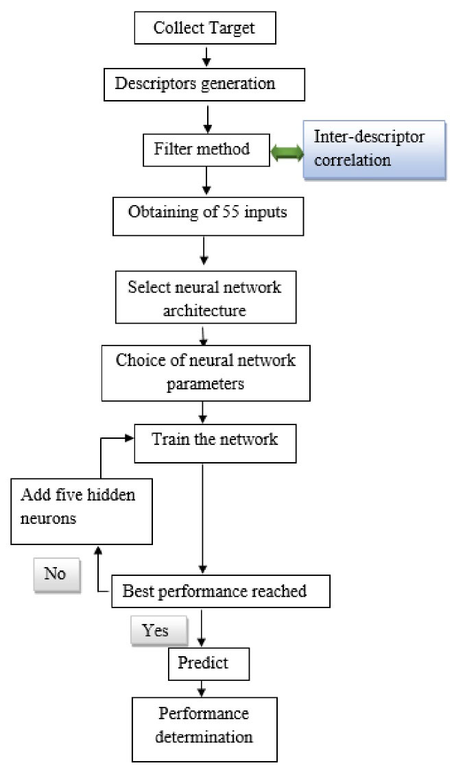
A five-step process was employed to predict the plasma protein-binding, as shown in Figure 1: (1) data set collection, (2) molecular descriptors generation, (3) selection of relevant descriptors by a filter method, (4) FFNN modeling, (5) validation of models.

Figure 1.
Flow sheet of the procedure followed
.
Flow sheet of the procedure followed
Data set collection
The experimental data values of protein-binding of the 277 drugs used in this study were selected from the pharmacological basis of the therapeutics handbook21 and the handbook of clinical drug data.22 Chemical names and experimental protein-binding values are presented in Supplementary file 1. This dataset was divided into two parts. The first one with 235 plasma protein-binding values, dedicated to develop the QSAR model. The second included 42 elements left for the external validation. The data was partitioned using holdout cross-validation.
Molecular descriptors generation
The numerical representation of molecular structure was assessed in terms of molecular descriptors; The SMILES script (simplified molecular input line-entry system) required to calculate descriptors was extracted from the open-access database PubChem.23 SMILES is a standard for specifying the structure of chemical species that takes the form of a line notation.24 Table 1 lists 1666 descriptors that were sorted into twenty categories using the SMILES scripts for the 277 drugs. The E-Dragon online programs,25 also known as the electronic remote version of the well-known software DRAGON created by the Milano Chemometrics and QSAR Research Group by Prof. R. Todeschini, were used to collect all descriptors. In Supplementary file 2, the name and number of calculated descriptors are presented.
Table 1.
Number of calculated descriptors and their categories
|
Descriptors category
|
Number
|
| Constitutional descriptors |
48 |
| Topological descriptors |
119 |
| Walk and pathcounts |
47 |
| Connectivity indices |
33 |
| Information indices |
47 |
| 2D autocorrelations |
96 |
| Edgeadjacency indices |
107 |
| Burdeneigen value descriptors |
64 |
| Topological charge indices |
21 |
| Eigen value based indices |
44 |
| Randic molecular profiles |
41 |
| Geometrical descriptors |
74 |
| RDF descriptors |
150 |
| 3D-morse descriptors |
160 |
| WHIM descriptors |
99 |
| GETAWAY descriptors |
197 |
| Functional group counts |
154 |
| Atomcentred fragments |
120 |
| Charge descriptors |
14 |
| Molecularproperties |
31 |
|
Total
|
1666
|
Selection of relevant descriptors
Feature selection techniques are applied to decrease the number of elements in the dataset by choosing features that will give us better accuracy with less data.26-28 It also reduces the overfitting and the overtraining risk.29 Feature selection methods are widely available in the literature. The characteristics, advantages, and disadvantages of the three main strategies that can be used for the selection of relevant descriptors are reported in Table 2.30
Table 2.
Feature selection methods and their advantages and disadvantages
|
Feature selection with filter methods
|
Feature selection with wrapper methods
|
Feature selection with embedded methods
|
| Relevance of the features is calculated by considering the intrinsic properties of the data. |
Wrapper methods select a subset of relevant features using a learning algorithm. |
Includes the classifier construction for the optimal feature selection. |
| Use feature relevance score to select the top rank features. |
Conduct search in the space of possible parameters. |
Like wrapper approaches, these methods are specific to a given learning algorithm. |
|
Examples
|
Examples
|
Examples
|
Information gain
Correlation coefficient scores
Chi squared test
T-test |
Genetic search
Sequential forward selection
Sequential backward elimination |
Decision tree
Weighted Naive Bayes
SVM |
|
Advantages
|
Advantages
|
Advantages
|
Can scale to high-dimensional data sets
Fast and computationally
inexpensive in comparison to wrapper method |
Considers features dependencies
Interaction with classifier
Simple to implement |
Classifier interaction
Considers feature dependencies |
|
Disadvantages
|
Disadvantages
|
Disadvantages
|
No interaction with the classifier
Univariate feature selection methods do not consider feature dependencies/ redundancy |
Higher risk of overfitting
Selection based on classifier
Computationally intensive |
Classifier dependencies |
The following procedure was used to reduce the number of molecular descriptors31:
simple
-
1. Descriptors having constant values (min = max) were eliminated.
-
2. Quasi-constant descriptors (1st quartile 25% = 2nd quartile 75%) were removed.
-
3. Descriptors with standard relative deviation RSD < 0.05 were deleted.
The three steps above were performed using STATISTICA software.32
simple
-
4. Matrices of the pairwise linear correlation between each pair of the column in the input matrices were calculated via MATLAB.33 Additionally, every variable that has a correlation coefficient R > 0.75 were removed. For more robustness of the model, the variance inflation factor VIF whose equation is as follows was calculated:
Where
is the squared correlation coefficient between the ith descriptor and the others. All descriptors with VIF > 5 were eliminated from the model.34
Model development
For the purpose of predicting the plasma protein-binding, the selected descriptors were used as inputs in FFNN. There are different approaches to discover the number of hidden neurons required for a modeling task explained in detail in a review named methods of selecting the number of hidden nodes in Artificial Neural Networks review.35 In this work, the following steps were used to choose the number of neurons in the hidden layer36:
simple
-
1. Initially, only five hidden neurons were taken.
-
2. The FFNN is trained until the mean square error does no longer seem to improve.
-
3. At this moment, five neurons are added to the hidden layer, each with randomly initialized weights, and resumed training.
-
4. The steps 2 and 3 are repeated until a termination criterion has been satisfied.
The mathematical equation of the model used for the prediction of protein binding is:
(2)
xi (i = 1…p) is the input that corresponds to the number of data included in the training of the ANN, i from 1 to 15, wij(i = 1…p, j = 1…k) are weights from input to hidden layer, b j (j = 1…k) are biases of the neurons in the hidden layer, k = 40 for filter method, w2j(j = 1…k) are weights from the hidden to the output layer, b is the bias of the output neuron and fb is the output.
Model validation
We established internal and external validation criteria to assess the QSAR models’ generalizability and predictive power. The following statistical parameters were used in our investigation to evaluate the models’ efficacy: the mean squared error (MSE), correlation coefficient (R), predictive squared correlation coefficient (Q2), and coefficient of determination (R2) values.
The residual sum of squares (RSS) is the difference between the fitted values and the observed values. The sum of squares (SS) refers to the difference between the observation and their mean. The PREdictive residual SS (PRESS) is the difference between the predictions and the observations.
Results and Discussion
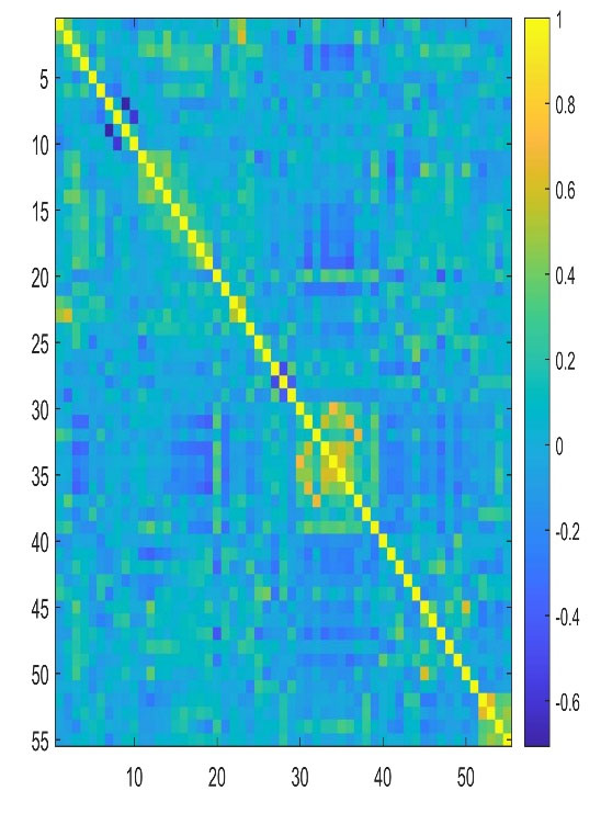
The results obtained from the selection of the most important descriptors using the correlation coefficient R and the variance inflation factor VIF showed that 55 descriptors seemed to be the most appropriate. The calculated VIFs among the values of the selected descriptors are less than five, indicating that multicollinearity between the selected descriptors is acceptable. To get an overview of the correlation structure we used a heatmap to highlight what is important (Figure 2). Table 3 shows the VIF values for the selected descriptors and their meanings.

Figure 2.
Heatmap of the correlation matrix for Filter method
.
Heatmap of the correlation matrix for Filter method
Table 3.
The VIF values for the selected descriptors by filter method
|
Descriptor
|
Type
|
Description
|
VIF
|
| nX |
Constitutional descriptors |
number of halogen atoms maximal |
2.6615 |
| MAXDN |
Topological descriptors |
Electrotopological negative variation |
4.4004 |
| MAXDP |
Topological descriptors |
maximal electrotopological positive variation |
3.5133 |
| PJI2 |
Topological descriptors |
2D Petitjean shape index |
1.7543 |
| Lop |
Topological descriptors |
Lopping centric index |
1.9696 |
| MATS1m |
2D autocorrelations |
Moran autocorrelation - lag 1 / weighted by atomic masses |
2.9735 |
| MATS2m |
2D autocorrelations |
Moran autocorrelation - lag 2 / weighted by atomic masses |
4.0324 |
| MATS4m |
2D autocorrelations |
Moran autocorrelation - lag 4 / weighted by atomic masses |
2.6631 |
| GATS2m |
2D autocorrelations |
Geary autocorrelation - lag 2 / weighted by atomic masses |
3.3624 |
| GATS4m |
2D autocorrelations |
Geary autocorrelation - lag 4 / weighted by atomic masses |
2.7846 |
| JGI2 |
Topological charge indices |
Mean topological charge index of order2 |
2.4942 |
| JGI3 |
Topological charge indices |
Mean topological charge index of order3 |
3.5132 |
| JGI4 |
Topological charge indices |
Mean topological charge index of order4 |
1.8913 |
| JGI5 |
Topological charge indices |
Mean topological charge index of order5 |
2.3317 |
| JGI6 |
Topological charge indices |
Mean topological charge index of order6 |
1.9848 |
| JGI7 |
Topological charge indices |
Mean topological charge index of order7 |
2.0805 |
| JGI8 |
Topological charge indices |
Mean topological charge index of order8 |
1.6600 |
| JGI9 |
Topological charge indices |
Mean topological charge index of order9 |
2.0077 |
| JGI10 |
Topological charge indices |
Mean topological charge index of order10 |
1.8634 |
| FDI |
Geometrical descriptors |
Folding degree index |
2.7337 |
| PJI3 |
Geometrical descriptors |
3D Petitjean shape index |
1.8725 |
| DISPm |
Geometrical descriptors |
d COMMA2 value / weighted by atomic masses |
2.3418 |
| DISPe |
Geometrical descriptors |
d COMMA2 value / weighted by atomic Sanderson electronegativities |
4.0247 |
| Mor04m |
3D-MoRSE descriptors |
3D-MoRSE - signal 04 / weighted by atomic masses |
2.4145 |
| Mor12m |
3D-MoRSE descriptors |
3D-MoRSE - signal 12 / weighted by atomic masses |
3.0770 |
| Mor17m |
3D-MoRSE descriptors |
3D-MoRSE - signal 17 / weighted by atomic masses |
1.8941 |
| Mor26m |
3D-MoRSE descriptors |
3D-MoRSE - signal 26 / weighted by atomic masses |
2.3693 |
| Mor28m |
3D-MoRSE descriptors |
3D-MoRSE - signal 28 / weighted by atomic masses |
2.5970 |
| Mor31m |
3D-MoRSE descriptors |
3D-MoRSE - signal 31 / weighted by atomic masses |
2.7935 |
| G2u |
WHIM descriptors |
2st component symmetry directional WHIM index / unweighted |
2.5765 |
| G2m |
WHIM descriptors |
2st component symmetry directional WHIM index / weighted by atomic masses |
2.7232 |
| E2m |
WHIM descriptors |
2nd component accessibility directional WHIM index / weighted by atomic masses |
2.9830 |
| G2v |
WHIM descriptors |
2st component symmetry directional WHIM index / weighted by atomic van der Waals volumes |
2.6210 |
| G2e |
WHIM descriptors |
2st component symmetry directional WHIM index / weighted by atomic Sanderson electronegativities |
3.5147 |
| G2p |
WHIM descriptors |
2st component symmetry directional WHIM index / weighted by atomic polarizabilities |
2.8308 |
| G2s |
WHIM descriptors |
2st component symmetry directional WHIM index / weighted by atomic electrotopological states |
2.9183 |
| E2s |
WHIM descriptors |
2nd Component accessibility directional WHIM index / weighted by atomic electrotopological states |
3.0706 |
| ISH |
GETAWAY descriptors |
Standardized information content on the leverage equality |
1.7718 |
| HATS4m |
GETAWAY descriptors |
Leverage-weighted autocorrelation of lag 4 / weighted by atomic masses |
3.2173 |
| C-005 |
Atom-centred fragments |
Atom-centred fragments |
2.0372 |
| C-006 |
Atom-centred fragments |
Atom-centred fragments |
2.3826 |
| C-008 |
Atom-centred fragments |
Atom-centred fragments |
2.7744 |
| C-025 |
Atom-centred fragments |
Atom-centred fragments |
2.4806 |
| C-026 |
Atom-centred fragments |
Atom-centred fragments |
3.2520 |
| C-040 |
Atom-centred fragments |
Atom-centred fragments |
3.5563 |
| H-048 |
Atom-centred fragments |
Atom-centred fragments |
1.9890 |
| H-052 |
Atom-centred fragments |
Atom-centred fragments |
2.3870 |
| O-057 |
Atom-centred fragments |
Atom-centred fragments |
2.2844 |
| O-060 |
Atom-centred fragments |
Atom-centred fragments |
2.7103 |
| N-072 |
Atom-centred fragments |
Atom-centred fragments |
2.5844 |
| N-075 |
Atom-centred fragments |
Atom-centred fragments |
2.0612 |
| Inflammat-80 |
Molecularproperties |
Ghose-Viswanadhan-Wendoloskiantiinflammatory at 80% (drug-like index) |
2.6589 |
| Hypertens-80 |
Molecularproperties |
Ghose-Viswanadhan-Wendoloski antihypertensive at 80% (drug-like index) |
2.8495 |
| Hypnotic-80 |
Molecular properties |
Ghose-Viswanadhan-Wendoloski hypnotic at 80% (drug-like index) |
2.4199 |
| Neoplastic-50 |
Molecular properties |
Ghose-Viswanadhan-Wendoloski antineoplastic at 50% (drug-like index) |
1.8707 |
We followed the above-mentioned procedure to determine the required number of hidden neurons. The best model’s accuracy was assessed using the R(all), MSE(validation),
, and Q2 criteria. The best model was chosen based on the maximum R(all),
, and Q2 and the lowest MSE (validation).31,37 Table 4 shows 10 network models developed. The results obtained show that network eight with 40 neurons is the best model with R (all) = 0.990,
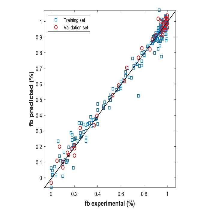
= 0.981, Q2 = 0.989, and MSE (validation) = 0.002. The best performance of the model had a topology of (55-40-1): 55 input nodes, one hidden layer with 40 nodes having the hyperbolic tangent as a transfer function, and one output layer with an identity function. The neural networks were implemented using Neural Network Toolbox for MATLAB.33 Figure 3 shows the predicted protein-binding values versus the experimental ones for the training and validation sets. The results show a close correlation between predicted and observed plasma protein-binding. The network type used is a Feed-Forward Network with the Levenberg-Marquardt backpropagation training function and gradient descent with momentum weight and bias learning function and the data was partitioned using holdout cross-validation. The difference between
and Q2 was equal to 0.008. this difference did not exceed 0.3 indicating the robustness of the model.38
Table 4.
Selected criteria of the different multi-layer perceptron for Filter method
|
Number of hidden neurons
|
R
(all)
|
|
Q2
|
MSE (validation)
|
| 5 |
0.849 |
0.743 |
0.707 |
0.039 |
| 10 |
0.857 |
0.729 |
0.664 |
0.032 |
| 15 |
0.870 |
0.774 |
0.743 |
0.031 |
| 20 |
0.872 |
0.780 |
0.714 |
0.038 |
| 25 |
0.917 |
0.839 |
0.780 |
0.026 |
| 30 |
0.957 |
0.918 |
0.882 |
0.020 |
| 35 |
0.955 |
0.953 |
0.818 |
0.024 |
| 40 |
0.990 |
0.981 |
0.989 |
0.002 |
| 45 |
0.944 |
0.901 |
0.875 |
0.014 |
| 50 |
0.832 |
0.694 |
0.714 |
0.027 |

Figure 3.
Comparison between experimental and predicted values for training and validation sets
.
Comparison between experimental and predicted values for training and validation sets
In order to investigate the predictability and performance of the model developed in this work, a statistical evaluation is carried out, as shown in Table 5. The model’s robustness is demonstrated by the fact that the internal validation’s statistical coefficients are all acceptable and satisfactory (lowest MSE, RMSE, and MAE, as well as high
, Q2,
). External validation parameters were also used to evaluate the model’s quality. We can say that this model stands out due to its high predictive power. The excellent Q2 value is greater than 0.9.38
Table 5.
External and internal criteria of the model
|
Parameters
|
Value
|
| Internal validation |
|
| R (all) |
0.991 |
|
|
0.981 |
|
Q2
|
0.989 |
| MSE |
0.002 |
| MAE |
0.028 |
| RMSE |
0.039 |
|
|
0.989 |
| External validation |
|
| R |
0.983 |
|
Q2
|
0.966 |
| MSE |
0.004 |
| MAE |
0.042 |
| RMSE |
0.063 |
Comparison between models from literature
We made a comparison between the few models reported in the literature with our developed model for the prediction of the binding of drugs to plasma proteins (Table 6). The evaluation of the advantages and disadvantages of these methods is quite difficult (each study used different data sets and different modeling approaches). We can see that the statistical parameters of our study exceed the models published previously. Our model gives a high R2, Q2,
and lowest MSE, RMSE, MAE. According to these results, our model can be used for predicting plasma protein binding for new drugs saving amounts of money and time.
Table 6.
Comparison with literature
|
Method
|
MAE
|
R2
|
R
|
MSE
|
| Suggested method (Filter method) |
Train 0.0313
Validation 0.0284
Test 0.0423 |
Train 0.981
Validation 0.989
Test 0.966 |
Train 0.991
Validation 0.995
Test 0.983 |
Train 0.031
Validation 0.028
Test 0.042 |
| Yuan et al18 |
Test 0.076 |
|
|
|
| Sun et al16 |
Test 0.126 |
|
|
|
| Kumar et al17 |
|
|
|
Train 0.869
Test 0.8881 |
| Li et al39 |
|
Train 0.86 |
|
|
| Ghafourian et al12 |
Train 13.25
Validation 14.96 |
Train 0.717
Validation 0.646 |
Train 0.681
Validation 0.641 |
|
| Moda et al40 |
|
Test 0.91 |
|
|
Applicability domain
A clearly defined applicability domain is recommended as the principle in OECD41 guidelines. In this work, we analyzed the domain of applicability with different approaches reported in Table 7 with the results. The proposed approaches’ algorithm and method can be found in the literature.42,43
Table 7.
Applicability domain for Filter method
|
Approach
|
Test inside AD
|
Test outside AD
|
| Bounding box (PCA) |
39 |
3 |
| Euclidan distance (95 percentile) |
40 |
2 |
| Classical KNN (Euclidean distance, k = 5) |
40 |
2 |
| KNN (Euclidean distance, k = 25) |
41 |
1 |
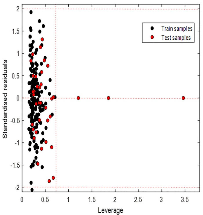
The number of samples inside the applicability domain varied depending on the method used. Euclidean distance (95 percentile) and Classical KNN (Euclidean distance, k = 5) identified two test samples out of the domain of applicability. KNN (Euclidean distance k = 25) showed one of the test samples out of the applicability domain. Bounding box considered 03 test samples out of the applicability domain as shown in Figure 4. Although our points are far from the rest of the observations, they are close to the regression fitted line because they have a small residual, we speak of good leverage points. These results show that the model can be used to predict plasma protein binding for new compounds that have not been tested.

Figure 4.
Plot of the residuals for calculated values of fb versus their experimental values for training and test sets
.
Plot of the residuals for calculated values of fb versus their experimental values for training and test sets
Conclusion
In this study, we constructed a QSAR model to predict 277 human plasma protein binding. The feature selection strategy by a Filter method has produced 55 inputs, which were used to train a FFNN for predictions. Examination of the estimates of external and internal criteria indicated that the QSAR model developed is robust, externally predictive, and distinguished by a good applicability domain. The external accuracy of the validation set was calculated by the Q2 and RMSE which are equal to 0.966 and 0.063 respectively. 98.30% of the external validation set is correctly predicted. According to the OECD principle, we can say that this QSAR model can be used to predict the fraction of human plasma protein binding for drugs that have not been tested to avoid chemical synthesis and reduce expansive laboratory tests.
Competing Interests
None to be declared.
Ethical Approval
Not applicable.
Supplementary Files
Supplementary file 1. Chemical names and experimental protein-binding values.
(pdf)
Supplementary file 2. The name and number of calculated descriptors.
(pdf)
References
- Luscombe D, Nicholls PJ. Processes of drug handling by the body. In: Smith HJ, ed. Smith and Williams’ Introduction to the Principles of Drug Design and Action. 3rd ed. Overseas Publishers Association; 1998. p. 1-31.
- Schmidt S, Gonzalez D, Derendorf H. Significance of protein binding in pharmacokinetics and pharmacodynamics. J Pharm Sci 2010; 99(3):1107-22. doi: 10.1002/jps.21916 [Crossref] [ Google Scholar]
- Zhivkova ZD. Quantitative structure - pharmacokinetics relationships for plasma protein binding of basic drugs. J Pharm Pharm Sci 2017; 20(1):349-59. doi: 10.18433/j33633 [Crossref] [ Google Scholar]
- Singh J, Petter RC, Baillie TA, Whitty A. The resurgence of covalent drugs. Nat Rev Drug Discov 2011; 10(4):307-17. doi: 10.1038/nrd3410 [Crossref] [ Google Scholar]
- Trainor GL. The importance of plasma protein binding in drug discovery. Expert Opin Drug Discov 2007; 2(1):51-64. doi: 10.1517/17460441.2.1.51 [Crossref] [ Google Scholar]
- Bohnert T, Gan LS. Plasma protein binding: from discovery to development. J Pharm Sci 2013; 102(9):2953-94. doi: 10.1002/jps.23614 [Crossref] [ Google Scholar]
- Lin X, Li X, Lin X. A review on applications of computational methods in drug screening and design. Molecules 2020; 25(6):1375. doi: 10.3390/molecules25061375 [Crossref] [ Google Scholar]
- Sliwoski G, Kothiwale S, Meiler J, Lowe EW Jr. Computational methods in drug discovery. Pharmacol Rev 2014; 66(1):334-95. doi: 10.1124/pr.112.007336 [Crossref] [ Google Scholar]
- Guo Y, Yu L, Wen Z, Li M. Using support vector machine combined with auto covariance to predict protein-protein interactions from protein sequences. Nucleic Acids Res 2008; 36(9):3025-30. doi: 10.1093/nar/gkn159 [Crossref] [ Google Scholar]
- Zhao C, Zhang H, Zhang X, Zhang R, Luan F, Liu M. Prediction of milk/plasma drug concentration (M/P) ratio using support vector machine (SVM) method. Pharm Res 2006; 23(1):41-8. doi: 10.1007/s11095-005-8716-4 [Crossref] [ Google Scholar]
- Zsila F, Bikadi Z, Malik D, Hari P, Pechan I, Berces A. Evaluation of drug-human serum albumin binding interactions with support vector machine aided online automated docking. Bioinformatics 2011; 27(13):1806-13. doi: 10.1093/bioinformatics/btr284 [Crossref] [ Google Scholar]
- Ghafourian T, Amin Z. QSAR models for the prediction of plasma protein binding. Bioimpacts 2013; 3(1):21-7. doi: 10.5681/bi.2013.011 [Crossref] [ Google Scholar]
- Turner JV, Maddalena DJ, Cutler DJ. Pharmacokinetic parameter prediction from drug structure using artificial neural networks. Int J Pharm 2004; 270(1-2):209-19. doi: 10.1016/j.ijpharm.2003.10.011 [Crossref] [ Google Scholar]
- Fu XC, Wang GP, Gao JQ, Zhan SY, Liang WQ. Prediction of plasma protein binding of cephalosporins using an artificial neural network. Pharmazie 2007; 62(2):157-8. doi: 10.1691/ph.2007.2.5735 [Crossref] [ Google Scholar]
- Deng L, Sui Y, Zhang J. XGBPRH: prediction of binding hot spots at protein-RNA interfaces utilizing extreme gradient boosting. Genes (Basel) 2019; 10(3):242. doi: 10.3390/genes10030242 [Crossref] [ Google Scholar]
- Sun L, Yang H, Li J, Wang T, Li W, Liu G. In silico prediction of compounds binding to human plasma proteins by QSAR models. ChemMedChem 2018; 13(6):572-81. doi: 10.1002/cmdc.201700582 [Crossref] [ Google Scholar]
- Kumar R, Sharma A, Siddiqui MH, Tiwari RK. Prediction of drug-plasma protein binding using artificial intelligence based algorithms. Comb Chem High Throughput Screen 2018; 21(1):57-64. doi: 10.2174/1386207321666171218121557 [Crossref] [ Google Scholar]
- Yuan Y, Chang S, Zhang Z, Li Z, Li S, Xie P. A novel strategy for prediction of human plasma protein binding using machine learning techniques. Chemometr Intell Lab Syst 2020; 199:103962. doi: 10.1016/j.chemolab.2020.103962 [Crossref] [ Google Scholar]
- Altae-Tran H, Ramsundar B, Pappu AS, Pande V. Low data drug discovery with one-shot learning. ACS Cent Sci 2017; 3(4):283-93. doi: 10.1021/acscentsci.6b00367 [Crossref] [ Google Scholar]
- Wallach I, Dzamba M, Heifets A. AtomNet: A Deep Convolutional Neural Network for Bioactivity Prediction in Structure-based Drug Discovery. arXiv [Preprint]. 10.48550/arXiv.1510.02855.
- Brunton LL, Knollmann BC. Goodman & Gilman’s: The Pharmacological Basis of Therapeutics. 12th ed. McGraw Hill; 2010.
- Anderson PO, Knoben JE, Troutman WG. Handbook of Clinical Drug Data. McGraw-Hill; 2002.
- National Institutes of Health (NIH). PubChem. Available from: https://pubchemdocs.ncbi.nlm.nih.gov/.
- Weininger D. SMILES, a chemical language and information system 1 Introduction to methodology and encoding rules. J Chem Inf Comput Sci 1988; 28(1):31-6. doi: 10.1021/ci00057a005 [Crossref] [ Google Scholar]
- Vcclab. Virtual Computational Chemistry Laboratory. 2005. Available from: http://www.vcclab.org/.
- Blum AL, Langley P. Selection of relevant features and examples in machine learning. Artificial Intelligence 1997; 97:245-271. doi: 10.1016/S0004-3702(97)00063-5 [Crossref] [ Google Scholar]
- Dash M, Liu H. Feature selection for classification. Intell Data Anal 1997; 1(1-4):131-56. doi: 10.1016/s1088-467x(97)00008-5 [Crossref] [ Google Scholar]
- Hira ZM, Gillies DF. A review of feature selection and feature extraction methods applied on microarray data. Adv Bioinformatics 2015; 2015:198363. doi: 10.1155/2015/198363 [Crossref] [ Google Scholar]
- Tetko IV, Livingstone DJ, Luik AI. Neural network studies. 1. Comparison of overfitting and overtraining. J Chem Inf Comput Sci 1995; 35(5):826-33. doi: 10.1021/ci00027a006 [Crossref] [ Google Scholar]
- Danishuddin Danishuddin, Khan AU. Descriptors and their selection methods in QSAR analysis: paradigm for drug design. Drug Discov Today 2016; 21(8):1291-302. doi: 10.1016/j.drudis.2016.06.013 [Crossref] [ Google Scholar]
- Hamadache M, Benkortbi O, Hanini S, Amrane A, Khaouane L, Si Moussa C. A quantitative structure activity relationship for acute oral toxicity of pesticides on rats: Validation, domain of application and prediction. J Hazard Mater 2016; 303:28-40. doi: 10.1016/j.jhazmat.2015.09.021 [Crossref] [ Google Scholar]
- Stat Soft. STATISTICA, version 8.0. Available from: https://www.statsoft.com/.
- MathWorks. MATLAB R2019b. Available from: https://www.mathworks.com/products/new_products/release2019b.html.
- Akinwande MO, Dikko HG, Samson A. Variance inflation factor: as a condition for the inclusion of suppressor variable (s) in regression analysis. Open J Stat 2015; 5(7):754-67. doi: 10.4236/ojs.2015.57075 [Crossref] [ Google Scholar]
- Panchal FS, Panchal M. Review on methods of selecting number of hidden nodes in artificial neural network. Int J Comput Sci Mob Computing 2014; 3(11):455-64. [ Google Scholar]
- Kubat M. An Introduction to Machine Learning. Cham, Switzerland: Springer; 2017.
- Bitam S, Hamadache M, Hanini S. QSAR model for prediction of the therapeutic potency of N-benzylpiperidine derivatives as AChE inhibitors. SAR QSAR Environ Res 2017; 28(6):471-89. doi: 10.1080/1062936x.2017.1331467 [Crossref] [ Google Scholar]
- Eriksson L, Jaworska J, Worth AP, Cronin MT, McDowell RM, Gramatica P. Methods for reliability and uncertainty assessment and for applicability evaluations of classification- and regression-based QSARs. Environ Health Perspect 2003; 111(10):1361-75. doi: 10.1289/ehp.5758 [Crossref] [ Google Scholar]
- Li H, Chen Z, Xu X, Sui X, Guo T, Liu W. Predicting human plasma protein binding of drugs using plasma protein interaction QSAR analysis (PPI-QSAR). Biopharm Drug Dispos 2011; 32(6):333-42. doi: 10.1002/bdd.762 [Crossref] [ Google Scholar]
- Moda TL, Montanari CA, Andricopulo AD. In silico prediction of human plasma protein binding using hologram QSAR. Lett Drug Des Discov 2007; 4(7):502-9. doi: 10.2174/157018007781788480 [Crossref] [ Google Scholar]
- OECD. Guidance Document on the Validation of (Quantitative) Structure-Activity Relationship [(Q)SAR] Models. Paris: OECD Publishing; 2014.
- Sahigara F, Mansouri K, Ballabio D, Mauri A, Consonni V, Todeschini R. Comparison of different approaches to define the applicability domain of QSAR models. Molecules 2012; 17(5):4791-810. doi: 10.3390/molecules17054791 [Crossref] [ Google Scholar]
- Roy K, Kar S, Ambure P. On a simple approach for determining applicability domain of QSAR models. Chemometr Intell Lab Syst 2015; 145:22-9. doi: 10.1016/j.chemolab.2015.04.013 [Crossref] [ Google Scholar]